


When fluid or infection creates a thick, restrictive layer around your lungs, breathing can become a struggle. This condition often requires intervention to help the lungs expand properly again. Fortunately, advancements in surgical techniques offer a less invasive solution than traditional open-chest procedures.
Video-Assisted Thoracoscopic Decortication (VATS) is a sophisticated surgical approach designed to address these complex lung conditions. It allows surgeons to skillfully remove the restrictive fibrous tissue, restoring lung function and improving your quality of life.
Table of Contents
- What is Video-Assisted Thoracoscopic Decortication (VATS)?
- When is VATS Decortication Necessary?
- Goals of VATS Decortication
- Preparing for Your VATS Procedure
- What to Expect During VATS Surgery
- Recovering After VATS Decortication
- Advantages of VATS Over Traditional Surgery
- Potential Risks and Complications
- Conclusion
What is Video-Assisted Thoracoscopic Decortication (VATS)?
Video-Assisted Thoracoscopic Decortication, often referred to as VATS decortication, is a minimally invasive surgical technique. Surgeons use this procedure to diagnose and treat various chest problems, specifically focusing on the lungs.
During VATS decortication, a surgeon removes a layer of fibrous tissue that covers the lung, chest wall, and diaphragm. This tough, constricting layer restricts the lung’s flexibility and prevents it from expanding and contracting effectively.
Unlike traditional open-chest surgery, which requires a large incision, VATS involves making several small incisions—typically three, each no more than 2.5 cm. These small openings allow the surgeon to insert a specialized miniature camera and surgical instruments into the chest cavity. The camera transmits live images from inside the chest to a video monitor, guiding the surgeon as they meticulously perform the decortication.
When is VATS Decortication Necessary?
Normally, a thin, fluid-filled space, less than 1 mm thick, exists between the lung and the chest wall. However, certain conditions can cause this space to fill with pus or blood, which can eventually solidify into a fibrous capsule around the lung, severely limiting its movement.
Empyema (Pleural Infection)
One of the primary reasons for VATS decortication is empyema, a type of infection where pus collects in the pleural space. Empyema is typically classified into three stages:
- Stage I: Often treated with antibiotics.
- Stage II: This stage commonly requires VATS decortication or fibrinolytics to break down the fibrous material.
- Stage III: Usually necessitates open-chest surgery due to extensive fibrosis.
VATS decortication is highly effective for Stage II empyema, removing the infection and allowing the lung to re-expand.
Other Conditions Requiring Decortication
Other health conditions that may necessitate VATS decortication include:
- Hemothorax: A collection of blood in the pleural space, often due to trauma, which can lead to fibrosis if not properly drained.
- Lung Tumors: In specific cases, VATS may be used to remove tumors or surrounding fibrous tissue.
- Inflammatory Chest Thickening: Conditions like rheumatoid arthritis can cause the pleura to thicken, restricting lung function.
Goals of VATS Decortication
The primary objectives of performing VATS decortication are clear and patient-focused:
- Restore Lung Function: Surgeons aim to remove the fibrous cell layer, allowing the lung to re-expand, regain its elasticity, and perform its vital function of taking in and expelling air.
- Eliminate Infection: For cases of empyema, the procedure effectively removes the source of infection, preventing its spread and recurrence.
- Prevent Deformity: By addressing the fibrosis early, VATS decortication helps prevent permanent lung deformity and long-term respiratory complications.
Preparing for Your VATS Procedure
Careful preparation is crucial to ensure the best possible outcome for VATS decortication. Your medical team will conduct several assessments:
Diagnostic Imaging and Tests
A computed tomography (CT) scan is a fundamental step before VATS decortication. It helps determine the stage and cause of the empyema or other pleural conditions. Additionally, your doctor may request other tests:
- Blood Tests: To assess overall health, blood clotting ability, and check for signs of infection.
- Lung Function Tests: To measure how well your lungs are working and gauge their capacity.
- Electrocardiogram (ECG): To evaluate your heart’s electrical activity and ensure it can withstand surgery.
Based on your specific health status, your surgeon will determine if you require any additional procedures or examinations.
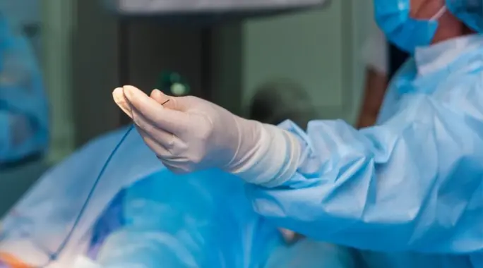
What to Expect During VATS Surgery
Understanding the surgical process can help ease anxieties. Here’s a general overview of what happens during VATS decortication:
- Anesthesia: You will receive general anesthesia before surgery, ensuring you are deeply asleep and pain-free throughout the procedure.
- Incisions: The surgeon will make several small incisions in your chest wall, typically three.
- Instrumentation: Through these incisions, the surgeon inserts a video camera (thoracoscope) and other specialized small surgical instruments.
- Procedure: Guided by the video feed, the surgeon meticulously removes the fibrous tissue and addresses any underlying issues.
- Closure: Once the medical procedure concludes, the surgeon removes all instruments, closes the incisions surgically, and covers them with bandages.
This procedure typically takes several hours to complete, depending on the complexity of the case.
Recovering After VATS Decortication
Your recovery journey begins immediately after surgery. Here’s what you can generally expect:
- Post-Anesthesia: You will typically wake up a few hours after surgery in a recovery room.
- Monitoring: Medical devices will monitor your vital signs, including heart rate, blood pressure, and oxygen levels.
- Oxygen Support: In some cases, you may receive supplemental oxygen through nasal tubes for a temporary period.
- Pain Management: It’s common to experience some pain, but it is usually manageable. You can request pain relievers from the medical staff.
- Chest Tube: A chest tube may be connected to help drain fluids or air from your lung, facilitating its re-expansion.
- Breathing Exercises: You will likely receive an incentive spirometer or similar device to help you breathe deeply, which is crucial for preventing lung infections.
- Blood Clot Prevention: Special compression stockings may be recommended to help prevent blood clots.
Most patients remain in the hospital for about 5-7 days until the chest tubes are removed and intravenous medications transition to oral ones. Your diet will gradually progress from liquids to solid foods as you recover.
Advantages of VATS Over Traditional Surgery
Compared to traditional open-chest surgery, VATS decortication offers significant benefits:
- Reduced Post-Operative Pain: The smaller incisions result in less pain after surgery, making recovery more comfortable.
- Shorter Hospital Stay: Patients often experience a quicker recovery, allowing for a shorter stay in the hospital.
- Faster Return to Activity: The minimally invasive nature of VATS generally leads to a faster recovery period and an earlier return to daily activities and work.
- Lower Risk of Complications: This technique reduces the risk of infections and excessive bleeding compared to more invasive procedures.
Potential Risks and Complications
While VATS decortication is generally safe, as with any surgical procedure, certain risks and potential complications exist:
- Lung Infection or Pneumonia: Despite preventive measures, there is a risk of developing a lung infection.
- Bleeding: Internal bleeding can occur during or after the surgery.
- Nerve Damage: Temporary or, rarely, permanent nerve damage in the chest area is a possibility.
- Organ Damage: Organs located near the surgical site could potentially be damaged.
- Anesthesia-Related Effects: Risks associated with general anesthesia, such as adverse reactions or respiratory issues, are always present.
Your surgical team will discuss these risks with you thoroughly before the procedure.
Conclusion
Video-Assisted Thoracoscopic Decortication (VATS) represents a vital advancement in treating complex lung and pleural conditions. This minimally invasive approach offers significant advantages, including less pain, faster recovery, and a reduced risk of complications, making it a preferred option for many patients. By removing restrictive fibrous tissue, VATS helps restore lung function and enhances overall quality of life, allowing individuals to breathe easier and live more actively.