
Finding out you have breast calcifications can be unsettling, but it’s important to remember that this is a common occurrence, especially for women over 50. While most breast calcifications are harmless and non-cancerous (benign), some types can indicate a higher risk of breast cancer. This often leads to a crucial question: Breast Calcifications: Do They Always Need a Biopsy?
Understanding when a biopsy is necessary, and when it’s not, can alleviate anxiety and help you navigate your breast health journey with confidence. Let’s explore what breast calcifications are, how doctors evaluate them, and what factors determine the need for further investigation.
- Understanding Breast Calcifications
- When Do Breast Calcifications Need a Biopsy?
- What to Expect During a Breast Biopsy
- Understanding Your Biopsy Results and Next Steps
- Conclusion: Prioritizing Your Breast Health
Understanding Breast Calcifications
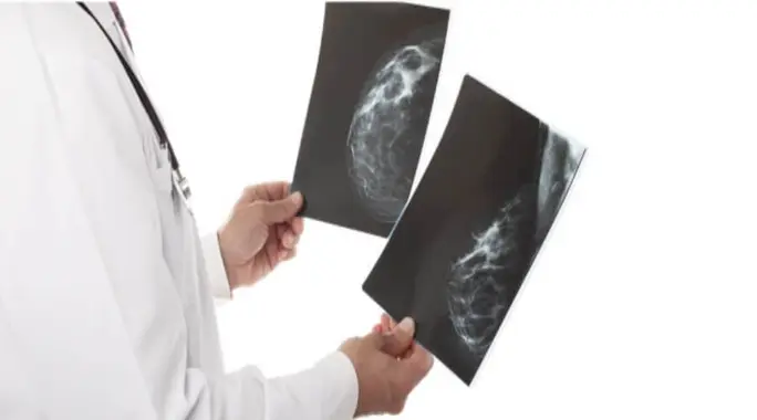
Breast calcifications are tiny calcium deposits that form within the breast tissue. They are so small that you cannot feel them during a self-exam. Instead, doctors usually detect them during a mammogram, appearing as bright white spots on the image.
These deposits are quite common, affecting about half of all women aged 50 and older. Their presence doesn’t automatically mean cancer; in fact, the vast majority are benign. However, radiologists carefully examine the size, shape, and pattern of these calcifications to determine their nature and potential significance.
When Do Breast Calcifications Need a Biopsy?
The good news is that if your breast calcifications are clearly identified as benign, a biopsy is typically not necessary. Your doctor will likely recommend routine follow-up mammograms to monitor them over time. However, a biopsy becomes crucial when calcifications appear “suspicious,” raising concerns about potential breast cancer.
A doctor will recommend a biopsy if there’s any doubt about the nature of the calcifications. This proactive step helps confirm whether they are benign or cancerous, guiding any necessary treatment decisions.
Key Characteristics of Suspicious Calcifications
Radiologists look for specific features that might suggest calcifications are cancerous. These characteristics often include:
- Small Size: Calcifications smaller than 5 millimeters (mm) can sometimes be a red flag.
- Irregular Shape and Size: Benign calcifications often look uniform. Irregularly shaped calcifications, or those varying significantly in size, warrant closer inspection.
- Linear or Clustered Patterns: When calcifications form a linear pattern, or are tightly grouped together in a specific area, it can indicate a higher risk.
If your mammogram shows any of these suspicious characteristics, your doctor will likely order additional imaging tests to get a clearer view.
The Diagnostic Process: From Mammogram to Biopsy Decision
After an initial mammogram reveals suspicious calcifications, your doctor will usually request a more detailed mammogram or other imaging studies. These advanced images allow for a better assessment of the calcifications’ nature and distribution.
If these further images confirm that the calcifications are concerning, your doctor will then recommend a breast biopsy. This procedure is the definitive way to determine if the cells are benign or cancerous, removing any uncertainty.
What to Expect During a Breast Biopsy
A breast biopsy is a common procedure used to collect a small tissue sample from the calcification area. This sample is then sent to a laboratory for microscopic examination. Here’s a general overview of the steps involved:
- You will lie comfortably on an examination table.
- The doctor will numb the breast area with a local anesthetic.
- Using imaging guidance (such as ultrasound, MRI, or stereotactic mammography), the doctor precisely locates the calcifications.
- A specialized needle is carefully inserted to extract one or more small tissue samples.
- Once the samples are collected, the doctor removes the needle, and a small dressing covers the insertion site.
- You might experience some mild pain or swelling in the biopsy area after the anesthetic wears off.
The laboratory typically takes a few days to several weeks to process the results, depending on the complexity of the analysis.
Understanding Your Biopsy Results and Next Steps
The steps following your biopsy depend entirely on the results:
- If the Calcifications are Benign: You can breathe a sigh of relief! No further treatment is usually needed. Your doctor will likely schedule a follow-up mammogram in about six months to ensure everything remains stable.
- If the Results Indicate Cancer: Your doctor will discuss the specific type of cancer detected and its extent. They will then develop a personalized treatment plan tailored to your needs. This plan might involve surgery, radiation therapy, chemotherapy, or a combination of treatments.
Regardless of the outcome, remember that early detection is a powerful tool for successful treatment. Do not hesitate to seek support from your medical team, family, and friends throughout this process.
Conclusion: Prioritizing Your Breast Health
While breast calcifications are a common finding, the critical distinction lies between benign and suspicious types. Most often, they are harmless and require no biopsy. However, if calcifications are small, irregular, or clustered, your doctor will recommend a biopsy to rule out cancer. This vigilant approach ensures that any potential issues are identified and addressed promptly, empowering you to take control of your breast health journey.